Challenges in the treatment of age-related macular degeneration
Macular degeneration
Vision impairment and blindness
Anti-VEGF agents have revolutionised outcomes for people with age-related macular degeneration (AMD) but nonadherence with the frequent injections needed and the monitoring required is a significant issue. New drugs are being developed that require fewer treatments, thereby improving treatment efficacy and reducing disease burden.
Age-related macular degeneration (AMD) is the leading cause of severe vision loss in our community and is the cause of blindness in about 50% of Australians who are legally blind.1 Intravitreally injected anti-vascular endothelial growth factor (anti-VEGF) drugs have significantly reduced blindness due to AMD complicated by choroidal bleeding. Unfortunately, the burden of continued monitoring and treatment and the economic costs associated with therapy for the patient and the Australian healthcare system have also increased significantly. Newer drugs and delivery systems and different treatment regimens are being investigated in the hope of being able to overcome these challenges.
It is estimated that in 2010 there were about one million Australians with AMD, equivalent to one in seven people over 50 years of age.1 Based on current projections, this will increase by over 70% to 1.77 million people by 2030.1 Twice as many patients will suffer bilateral visual impairment, up from 107,000 in 2010 to more than 215,000 by 2030.1
Clinical presentation and diagnosis
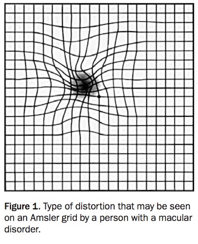
Patients with AMD often present to their GP with acute blurred vision or distorted central vision (metamorphopsia). Metamorphopsia may have been self-detected using an Amsler grid (Figure 1). Other symptoms may include central scotoma or decreased contrast sensitivity. Early ophthalmology referral is warranted when patients have developed any of these symptoms.
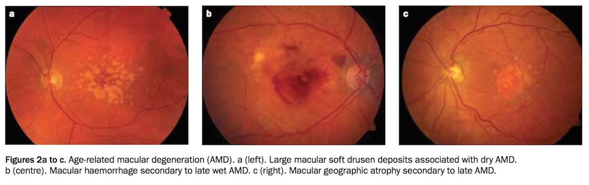
AMD may be classified into early ‘dry’ and late stages. In the early dry form, cellular debris (drusen) accumulates between the retina and the choroid and is associated with changes in the macular pigment (Figure 2a). Clinical progression is usually slow, and patients are often asymptomatic. There is currently no treatment available for early AMD and ongoing monitoring for late AMD is the mainstay of management. Dietary supplementation to slow clinical progression may be indicated in some forms of early AMD.
Late AMD can be divided into wet (neovascular) and dry (atrophic) forms. Wet AMD results from a growth of abnormal vasculature from the choroid (the vascular tissue layer beneath the retina) in response to vascular endothelial growth factor (VEGF) expressed by the disrupted retinal pigment epithelium.2 This process is termed choroidal neovascularisation. These vessels leak or bleed, causing serous or haemorrhagic elevation of the macula and eventual scarring (Figure 2b). This form is often associated with rapid, severe deterioration of vision, particularly if choroidal neovascularisation is active. Wet AMD is treatable with intravitreal injections of anti-VEGF agents (see below).
‘Late dry’ or atrophic AMD refers to the formation of geographic atrophy resulting from end-stage cell death of the retinal pigment epithelium and photoreceptors. These appear as pale islands at the macula (Figure 2c). Vision loss in atrophic AMD is variable depending on foveal involvement. When the fovea is involved, central vision loss is irreversible and dense. Because clinical progression is gradual, patients may learn to adapt. Unlike wet AMD, there is no treatment available for this form of advanced AMD.
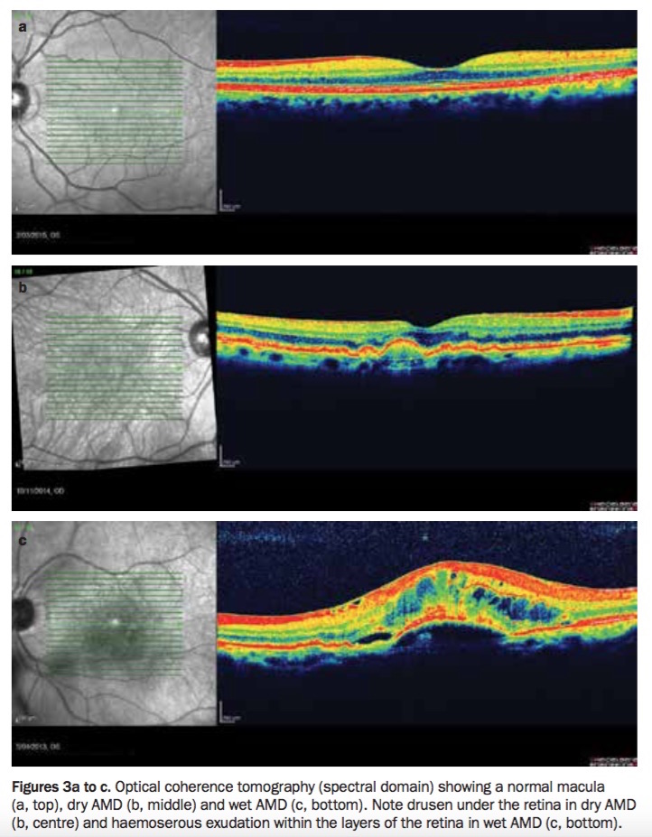
A diagnosis of dry AMD may be made based on macular changes seen on fundal examination alone. These changes may also be seen on optical coherence tomography (OCT), especially spectral domain OCT, a noninvasive imaging modality that provides a cross-sectional view of the retinal layers through the macula (Figures 3a to c).
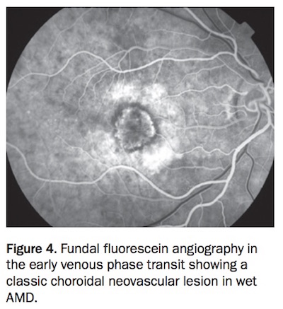
The standard diagnostic investigation in wet AMD is fundal fluorescein angiography (FFA) to identify choroidal neovascularisation if it is present (Figure 4). FFA is a test that involves taking serial images of the fundus after an intravenous injection of fluorescein dye and looking for choroidal neovascular lesions. OCT can be used in patients with wet AMD to identify subretinal fluid and retinal oedema resulting from active choroidal neovascularisation (Figure 3c).
Risk factors and prevention
The main risk factors for the development of advanced AMD are increasing age, ethnicity and genetics.3-6 Cigarette smoking is the main modifiable risk factor that has been consistently identified in numerous studies, showing a direct correlation of relative risk with number of pack years smoked.3,6 Cessation of smoking is strongly recommended as part of AMD management. Other modifiable risk factors, including body mass index, cardiovascular disease and hypertension, have been inconsistently associated with AMD incidence in population-based studies.5
Several studies have investigated the effects of diet on the progression of AMD. The Age-Related Eye Disease Study (AREDS) examined the potential benefits of high-dose oral antioxidants (vitamin C, vitamin E, beta-carotene) and zinc supplementation in reducing AMD progression, and showed a reduction in AMD progression of 25%.5,7 The AREDS2 study examined the same outcome using lutein/zeaxanthin instead of beta-carotene, and showed a similar reduction in AMD progression.8 Dietary fat intake has also been investigated. The AREDS study findings suggested high omega-3 long-chain polyunsaturated fatty acid consumption (from sources such as fish) reduced AMD incidence by 30% at 12 years.7 Despite this dietary association, AREDS2 failed to demonstrate a significant benefit from omega-3 polyunsaturated fatty acid oral supplementation.8
An increased risk of AMD has been found in individuals who have a higher intake of saturated fats and cholesterol and in those with a higher body mass index in population studies.5
Anti-vascular endothelial growth factor agents
A decade ago, wet AMD was considered untreatable and 60% of patients were expected to be legally blind within two years of diagnosis.1 Today, anti-VEGF agents have revolutionised AMD outcomes and have superseded treatments such as thermal laser photocoagulation (‘hot laser’) and nonthermal laser photodynamic therapy (‘cold laser’), with better long-term vision outcomes and lower complication rates.9-12
There are currently three anti-VEGF agents in clinical use: ranibizumab, aflibercept and bevacizumab (off-label use). They have been shown to be effective in treating choroidal neovascularisation in AMD in several clinical trials.13-17
Delivery
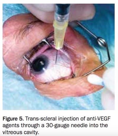
Anti-VEGF agents are administered by trans-scleral injection through a 30-gauge needle into the vitreous cavity using strict aseptic technique and usually under local anaesthesia (Figure 5).
Transient symptoms of discomfort, superficial haemorrhage on the conjunctiva and floaters related to disruption of the vitreous gel are common after an injection. Serious complications include intraocular infection, traumatic cataract, retinal detachment and haemorrhage into the vitreous cavity.
Systemic risks
Systemic anti-VEGF treatment carries theoretical risk of systemic arterial thromboembolic events. Clinical trials of intravitreal anti-VEGF injections have not shown this to be a significant concern. The VEGF Trap-Eye: Investigation of Efficacy and Safety in Wet AMD (VIEW) study, which examined the efficacy of aflibercept for choroidal neovascularisation, showed no statistically significant differences in rates of mortality and arteriothrombotic or venous-thrombotic events between ranibizumab and aflibercept.13 Similarly, the Comparison of Age-Related Macular Degeneration Treatments Trials (CATT) study did not show statistically significant differences between bevacizumab and ranibizumab at one year.14
The safety profile in pregnant or lactating women has not yet been established.18
Adherence and the burden of therapy
Clinical trials have provided strong evidence supporting monthly use of intravitreal injections, particularly with aggressive choroidal neovascular lesions. This burden of clinic visits and injections is significant for patients and their carers, often resulting in discontinuation of therapy. Clinical guidelines are inconsistent with regard to how long treatment needs to be continued.
Nonadherence with anti-VEGF therapy is a significant issue. It has been shown to be associated with poorer visual outcomes and higher risk of disease recurrence.19 Observational data for Australian patients on anti-VEGF treatment demonstrated a 42% discontinuation rate over a six-year period.20 Contributing factors for lack of adherence include treatment intolerance, health literacy, costs, perceived treatment necessity and perceived drug efficacy.19,20
Future treatments for AMD
New drugs targeting various pathways involved in the pathogenesis of choroidal neovascularisation are now undergoing phase II or phase III clinical trials. Most of these novel drugs aim to improve efficacy and reduce disease burden by reducing the number of treatments needed.2 They include:
- designated ankyrin repeat proteins (DARPins), which target angiogenesis with VEGF-A antagonism and destabilise vessel formation by stripping pericytes (vascular smooth muscle cells)
- platelet-derived growth factor, which targets the complement pathway that has been implicated in choroidal neovascularisation formation2
- antibodies to sphingosine-1-phosphate (S1P), which is implicated in the production of VEGF, fibroblast growth factor, platelet derived growth factor and related growth factors involved in choroidal neovascular lesion pathogenesis.21
Novel methods being explored include intravitreal APL-2 (a complement inhibitor), which is currently in phase III trial stage for the treatment of geographic atrophy (ClinicalTrials.gov Identifier NCT02503332). Several phase I trials are also being conducted for stem cell and gene therapeutic approaches.22 Prototypes of implantable drug delivery devices, such as microelectromechanical systems, are in the preclinical phase of development. Similarly to insulin pumps, they provide a refillable drug reservoir as an alternative to repeated injections.
Conclusion
AMD is the leading cause of visual impairment in Australia. Early diagnosis and treatment with intravitreal injections of anti-VEGF drugs saves sight. The burden of such treatments may be challenging to patients and carers and may reduce adherence to therapy. Investigations of alternative therapeutic targets and modalities are providing promising results.
COMPETING INTERESTS: None.
References
- Deloitte Access Economics; Mitchell P. Eyes on the future: a clear outlook on age-related macular degeneration. Macular Degeneration Foundation. Sydney: Deloitte Access Economics; 2011.
- Nagineni CN, Kommineni VK, William A, Detrick B, Hooks JJ. Regulation of VEGF expression in human retinal cells by cytokines: implications for the role of inflammation in age-related macular degeneration. J Cell Physiol 2012; 227: 116-126.
- Delcourt C, Diaz JL, Ponton-Sanchez A, Papoz L. Smoking and age-related macular degeneration. The POLA Study. Pathologies Oculaires Liées á l’Age. Arch Ophthalmol 1998; 116: 1031-1035.
- Klein R, Knudtson MD, Lee KE, Gangnon RE, Klein BE. Age-period-cohort effect on the incidence of age-related macular degeneration: the Beaver Dam Eye Study. Ophthalmol 2008; 115: 1460-1467.
- McCarty CA, Mukesh BN, Fu CL, Mitchell P, Wang JJ, Taylor HR. Risk factors for age-related maculopathy: the Visual Impairment Project. Arch Ophthalmol 2001; 119: 1455-1462.
- Tan JS, Mitchell P, Kifley A, Flood V, Smith W, Wang JJ. Smoking and the long-term incidence of age-related macular degeneration: the Blue Mountains Eye Study. Arch Ophthalmol 2007; 125: 1089-1095.
- Age-Related Eye Disease Study Research Group. A randomized, placebo-controlled, clinical trial of high-dose supplementation with vitamins C and E, beta carotene, and zinc for age-related macular degeneration and vision loss: AREDS report no 8. Arch Ophthalmol 2001; 119: 1417-1436.
- Age-Related Eye Disease Study 2 Research Group. Lutein + zeaxanthin and omega-3 fatty acids for age-related macular degeneration: the Age-Related Eye Disease Study 2 (AREDS2) randomized clinical trial. JAMA 2013; 309: 2005-2015.
- Azab M, Benchaboune M, Blinder KJ, et al. Verteporfin therapy of subfoveal choroidal neovascularization in age-related macular degeneration: meta-analysis of 2-year safety results in three randomized clinical trials: Treatment of Age-Related Macular Degeneration with Photodynamic Therapy and Verteporfin in Photodynamic Therapy Study report no. 4. Retina 2004; 24: 1-12.
- Brown DM, Michels M, Kaiser PK, et al. Ranibizumab versus verteporfin photodynamic therapy for neovascular age-related macular degeneration: two-year results of the ANCHOR study. Ophthalmology 2009; 116: 57-65.
- Laser photocoagulation of subfoveal neovascular lesions of age-related macular degeneration: updated findings from two clinical trials. Macular Photocoagulation Study Group. Arch Ophthalmol 1993; 111: 1200-1209.
- Larsen M, Schmidt-Erfurth U, Lanzetta P, et al; MONT BLANC Study Group. Verteporfin plus ranibizumab for choroidal neovascularization in age-related macular degeneration: twelve-month MONT BLANC study results. Ophthalmology 2012; 119: 992-1000.
- Heier JS, Brown DM, Chong V, Korobelnik JF, et al. Intravitreal aflibercept (VEGF trap-eye) in wet age-related macular degeneration. Ophthalmology 2012; 119: 2537-2548.
- CATT Research Group, Martin DF, Maguire MG, et al. Ranibizumab and bevacizumab for neovascular age-related macular degeneration. N Engl J Med 2011; 364: 1897-1908.
- Kaiser P, Brown D, Zhang K, et al. Ranibizumab for predominantly classic neovascular age-related macular degeneration: subgroup analysis of first-year ANCHOR results. Am J Ophthalmol 2007; 144: 850-857.
- Mitchell P, Korobelnik J, Lanzetta P, et al. Ranibizumab (Lucentis) in neovascular age-related macular degeneration: evidence from clinical trials. Br J Ophthalmol 2010; 94; 2-13.
- Regilo CD, Brown DM, Abraham P, et al. Randomized, double-masked, sham-controlled trial of ranibizumab for neovascular age-related macular degeneration: PIER Study year 1. Am J Ophthalmol 2008; 145: 239-248.
- Tarantola RM, Folk JC, Boldt HC, Mahajan VB. Intravitreal bevacizumab during pregnancy. Retina 2010; 30: 1405-1411.
- McHorney CA, Spain CV. Frequency of and reasons for medication non-fulfillment and non-persistence among American adults with chronic disease in 2008. Health Expect 2011; 14: 307-320.
- Vaze A, Fraser-Bell S, Gillies M. Reasons for discontinuation of intravitreal vascular endothelial growth factor inhibitors in neovascular age-related macular degeneration. Retina 2014; 34: 1774-1778.
- Sabbadini R. Sphingosine-1-phosphate antibodies as potential agents in the treatment of cancer and age-related macular degeneration. Br J Pharmacol 2011; 162: 1225-1238.
- Nazari H, Zhang L, Zhu D, et al. Stem cell based therapies for age-related macular degeneration: the promises and the challenges. Prog Retin Eye Res 2015; 48: 1-39.